-
Professor Chun Heungjae Team at CUK College of Medicine, Developed a New Patch for Regenerative Cardiac TissueAuthor : 관리자Date : 2023.03.20Hit : 351
-
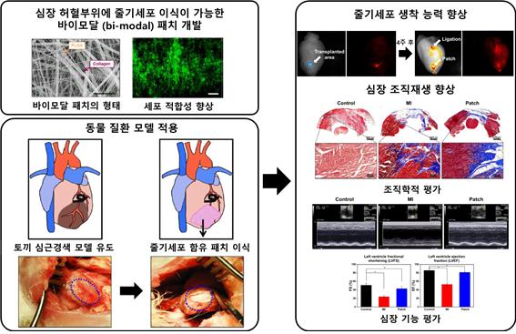
- Developed a new patch-type bi-modal scaffold composed of synthetic PLGA and nano/microfibers
- Extend the stem cell engraftment time from the existing 48 hours to over 4 weeks, significantly improving the heart functions
- Published in ""biomaterials research (IF:15.863),"" and selected and listed as ""People Who Made Korea Shine"" by the Biological Research Information Center(BRIC)
Professor Chun Heungjae (Director of the Cell & Tissue Engineering Institute) of the CUK College of Medicine created ""a patch to regenerate cardiac tissue"" through an interdisciplinary study with a clinical research team at the Catholic Medical Center.
Ischemic heart disease is caused by a lack of self-renewal ability of cardiomyocytes and is regarded as one of the leading causes of death worldwide, despite recent advances in drug and cutting-edge medical technology.
For the past 20 years, a number of basic and clinical tests using stem cells as alternative sources have been conducted in order to recover damaged cardiomyocytes. However, in the case of the heart, the engraftment rate of the transplanted stem cells is only around 5 to 10%, and even the transplanted stem cells have a low survival rate due to insufficient oxygen and nutrients, making the effectiveness of a cell therapy product significantly low.
To tackle the issue, a new strategy based on scaffold-based regenerative cardiac tissue engineering medicine has emerged. While the patch-type scaffold made of nanoscale fibers is in the spotlight, it also has limitations in that it makes it difficult for cells to freely infiltrate, which is required for stem cells to develop into a three-dimensional construct.
To address the issue, Professor Chun's research team created a new bi-modal patch made of collagen nanofibers, a component of natural extracellular matrix, and synthetic PLGA microfibers, which are widely used in surgical procedures. Professor Wee Junghee from Emergency Medicine (Yeouido St. Mary's Hospital), Professor Yoo Kidong from Cardiology (St. Vincent's Hospital), Professor Sim Sungbo Sim from Thoracic Surgery (Bucheon St. Mary's Hospital), and others reviewed the feasibility and applicability of the bi-modal patch as a cardiac tissue engineering material.
The bi-modal electrospinning scaffold developed by Professor Chun’s research team is a nano/micro bimodal composite fibrous patch composed of collagen and poly (D, L-lactic-co-glycolic acid) (Col/PLGA). It consists of an independent nozzle control multi-electrospinning apparatus, and its feasibility as a stem cell-laden cardiac patch was thoroughly investigated.
Nano/Micro bimodal distributions of Col/PLGA patches were obtained in the 4-6% collagen concentration range, while co-electrospinning improved collagen's poor mechanical properties and PLGA's hydrophobic properties. In vitro tests with bone marrow-derived mesenchymal stem cells (BMSCs) revealed that Col/PLGA had better cytocompatibility and proliferation capacity than PLGA, and their extent increased as collagen content increased.
The results of tracing nanoparticle-labeled and GFP(green fluorescent protein)-labeled BMSCs strongly support that Col/PLGA has the ability to retain stem cells for an extended period of time and therefore allow stem cells to directly affect myocardial and vascular endothelial cells or secrete recovery factors, leading to improved heart function as evidenced by histological and echocardiographic findings.
As a result of the enhanced cyto-compatibility and proliferation capacity demonstrated by bi-modal patches in vitro and in vivo experiments using BMSC, stem cell tracking using nanoparticle and GFP marks indicated that bi-modal patches could be engrafted for a long time over four weeks. Given that the normal engraftment period of transplanted cells based on current cell therapy methods is 48 hours, this is an undeniably impressive performance.
Furthermore, it was strongly suggested that the transplanted stem cells could directly affect myocardial and vascular endothelial cells or secrete recovery factors. As a result, histological and echocardiographic findings show that it significantly improves heart functions.
""This was an interdisciplinary project combining engineering, basic and clinical medicine, and despite difficulties in producing the results, we were able to obtain satisfactory results thanks to the proactive participation of researchers,"" said Professor Chun Heungjae. ""As a follow-up to this study, we will soon announce another study result that is more practical in nature and includes radiological findings for animals such as CT,"" he added.
The study, on the other hand, was recently chosen and listed as “People Who Made Korea Shine” by the Biological Research Information Center (BRIC) and published in one of the world's most prestigious journals, “Biomaterials research (IF:15.863)” under the title “Stem cell laden nano and micro collagen/PLGA bimodal fibrous patches for myocardial regeneration.” (References, Photo available) (end).
[References]
Reference 1.
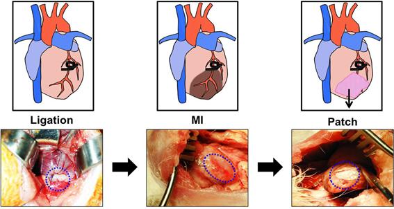
Reference 2. A cardiac patch is implanted in an ischemic section of a rabbit heart
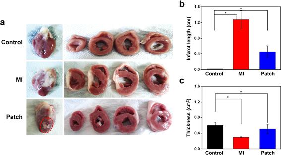
Reference 3. Regeneration and cardiac function improvement around the infarcted area using the cardiac patch
-
Attachment File

